FIS4-3D living cell microscope
Marker-free 3D living cell imagin
Near infrared, LED white wide spectrum lighting
Easy to integrate all types of microscopes, plug and play,
lightweight and compact

New products for biological and Medical cell culture research and testing: 3D living cell microscopy (marked free 3D real-time imaging) can make up for the shortcomings of phase contrast microscopy, fluorescence microscopy, confocal microscopy in 3D quantitative phase, in vivo, high efficiency and other aspects, realize cell biopsy, cell count, red blood cell pathological screening, cancer living tissue imaging, and provide quantitative analysis of cell contour information 3D morphology and quality!

High contrast
Displays 3D images of cells and various micro and nano fine structures

High efficiency
The detection system is stable and the cell image adjustment is convenient

Living cell detection
Non-staining, non-damaging, long-lasting biopsies of biological cells

Fully functional
With all the viewing functions of a conventional inverted microscope
◆ Real-time 3D morphologic imaging of living cells
◆ 4-D timeline - live cell culture, live cell diagnosis
◆ Live cell segmentation count, dry weight measurement
◆ Micro and nano structure measurement of transparent material
The mobile platform is fixed on the main body of the microscope by magnetic suction, and the electric and manually controlled mobile platform can be replaced according to needs.
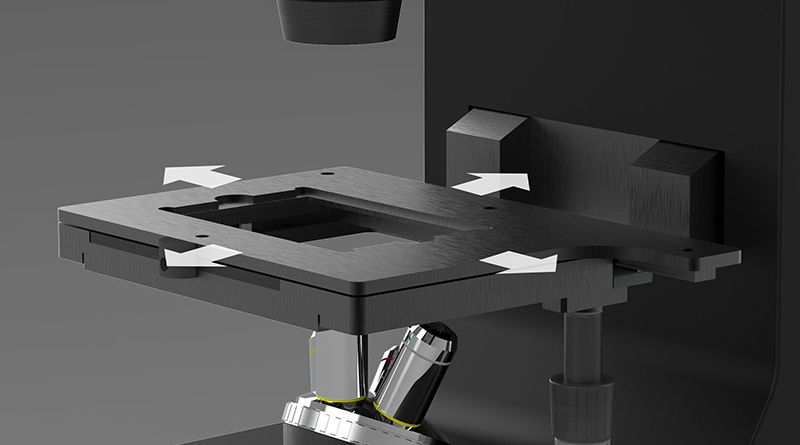
Manual control
Manual control stroke,X:190mm,Y:90mm
Table size:220mm×170mm

Electric control
Electric control stroke,X:110mm,Y:75mm
Table size:260mm×153mm

controller
Through the controller, the objective lens can be adjusted in the Z direction at low, medium and three speeds. The Z direction stroke is 30mm
By flipping the main body of the microscope and flipping the fixed moving platform, the 3D living cell microscope can be flipped to facilitate the magnification switching and observation in different occasions.

|
Model number |
FIS4-3DCell |
|
dimension |
355mm(L)×258mm(W)×437mm(H) |
|
X Y Travel range (manual) |
X:130mm,Y:90mm |
|
X Y Travel Range (electric) |
X:110mm,Y:75mm |
|
Focus Z-axis travel (electric) |
30mm |
|
test sample |
Biological cells - can be directly put into the petri dish test - viable, transparent material micro and nano structure |
|
CCD pixel |
2048*2048 |
|
Resolution (vertical and horizontal) |
2nm(longitudinal); 1μm(transverse) |
|
Wave length |
LED white light |
|
Microscope objective |
Options:5X、10X、20X、50X、100X |
|
Display frame rate in real time |
10HZ |
|
Software system |
Direct display output 3D images, data files |
|
Adaptive sensor |
FIS4-Cell |


INQUIRY


Add Successfully
 CONTACT

7X24小时电话 周一至周六 9:00-18:00
0757-6668-88268
Hangzhou Zernike Optical Technology Co., LTD
Full range of authorized dealers
Jiangsu Yucheng light sense technology Co., LTD
7X24 hours telephone Monday to Saturday 9:00-18:00
180-1280-4887

电话:0757-666888268
传真:0757-666999368
公司地址:佛山市禅城区某某某路同某某莫大厦1208
©2021 版权空气净化机租赁所有
备案号:粤ICP备100000000-1号
©2024 Copyright Jiangsu Yucheng Light Sense Technology Co., LTD
Record number: Su ICP for 2024068497
TEL:18012804887
EMAIL:zernikeoptics@zernikeoptics.cn
Address::No. 96-3 Tianpu Road, Jiangbei New District, Nanjing City, Jiangsu Province


点击右上角
分享给朋友吧